Showing posts with label amino acids. Show all posts

Sunday, June 15, 2014

The Basics of Protein Structure Part 1: The Levels of Structure

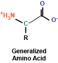
In a previous post, we had explored the characteristics of the 20 amino acids that make up proteins.  These amino acids make up a diverse collection of molecules that can be strung together, making up proteins that have a multitude of functions within the cell.

Amino acids form a protein through the action of the ribosome, which we will discuss in a future post.  At this time, suffice it to say that the ribosome uses an elegant mechanism to read mRNA and translate it into the protein encoded by the mRNA by adding amino acids in a string.  As this string of amino acids is created, it begins to form a structure that will have functions within the cell.

Four levels of protein structure exist:
Primary (1o) structure:  The order of the amino acids is the primary structure.  Think of the primary structure as the alphabet of the amino acids: MGRYNVPL, for example.  The primary structure describes what order amino acids are in, and even though it might not seem like very much information, even the primary structure of a protein can provide a great deal of information in terms of its function and its potential 3-dimensional shape.

Secondary (2o) structure:  When amino acids are polymerized, they form local structures, which make up the secondary structure.  Think of secondary structure as the shape of a group of amino acids.  Two primary forms of secondary structure exist:  alpha helices and beta sheets.  Alpha helices result from the coiling of the amino acid string turning about itself.  In contrast, beta sheets are flatter and lack coiling.  There are several types of alpha helices and beta sheets, which we will save for a future post, and these different types of structures have important implications for overall shape of a protein.

Tertiary (3o) structure:  Protein structure gets exciting when you talk about tertiary structure, which can be described as the overall three-dimensional shape of a protein.  In general, the tertiary structure is the “final” form of a protein, although modifications on the protein, as well as interactions with other proteins can affect this structure.


Quaternary (4o) structure:  When proteins interact with each other, they can form complexes, which is the quaternary structure of these proteins.  You can think of this structure as the way proteins contact each other.  The bundling of proteins together can be between proteins of the same type (such as is the case with hemoglobin) or other types of proteins.

The drawing attempts to illustrate the concept of the different levels of protein structure.  Again, think of the primary structure as the order; the secondary structure as the local shape; the tertiary structure as the overall shape; and the quaternary structure as the way this tertiary structure of the protein interacts with other proteins.

How protein structure is established is a fascinating question and a field that is actively studied by prominent labs around the world.  Protein folding is the process of a chain of amino acids curling into its final shape, and how this process occurs is complex and not completely understood.  In general proteins fold depending on their environment (exposed to water or not, for example) and with the help of other proteins, called chaperones.  Protein chaperones help to establish a protein’s structure as well as maintain it during times of stress.  Further, modifications on proteins can change their structures, such as when p53, a protein that is involved in regulating many processes within the cell, is phosphorylated – its structure and, consequently, its function is altered slightly. 

The structure of proteins has fascinated scientists since we first learned about proteins.  Thousands of scientists around the world are still working to discover new structures and to learn how proteins fold.  The science behind protein folding has important implications in diseases such as Alzheimer’s, cancers, and infectious diseases.   

Monday, October 17, 2011

The Trp Operon

The last post about an operon (the lac operon) is the most viewed post on this blog, so I thought that it might be helpful to follow this up with another operon, this time concentrating on the trp (tryptophan) operon.  This operon is another really elegant example of transcriptional regulation in E.coli and the mechanism is pretty cool.

Amino acids are essential for life (see the last post on their composition!) and cells synthesize amino acids using a variety of enzymes.  When nutrients are plentiful, such as E.coli would encounter in nutrient broth in the laboratory setting, cells no longer need to waste energy producing biosynthetic enzymes when they can utilize nutrients already in excess.  The trp operon contains several enzymes that are coordinately regulated and involved in the production of tryptophan.  When tryptophan is present in the cell's environment, it doesn't need to make any of these enzymes, but if the cell needs tryptophan, these enzymes are transcribed and shortly thereafter translated.  Control of this operon, thus, controls how much energy the cell is going to put into making tryptophan.

Similar to the lac operon, the trp operon contains an operator (O) sequence, within the promoter sequence, where an operator binds and prevents transcription.  In the presence of tryptophan, the operator binds the promoter and prevents RNA polymerase from transcribing genes.  In the absence of tryptophan, however, transcription occurs at a basal rate.  Sounds simple enough, right?  Let's take it a step further and consider...

Attenuation
An important concept in gene regulation is that of attenuation, which is fine-tuning of gene expression.  You might think that attenuation is mediated by protein factors that bind the DNA and affect gene expression; however, attenuation of the trp operon is a little different and, instead, depends on mRNA structure to modulate gene expression.

Before moving forward, let's look at the trp operon (diagrammed to the right).  Briefly,t here are four regions, and these four regions have differing levels of complementarity to each other.  Thus, when the DNA is transcribed into mRNA, the mRNA folds into all kinds of shapes and the regions of the trp operon fold on each other.

After transcription of the entire trp operon (we're dealing with mRNA from this point forward), the next event is translation of this mRNA into protein.  In bacteria, it's important to note that transcription and translation occur simultaneously, so as soon as we have a transcript in a bacterial cell, it's being translated.  The trp transcript contains two critical tryptophan codons immediately before region 1, so in order to synthesize the enzymatic machinery to make tryptophan, the cell must use a few residues to translate the protein.

In the presence of high amounts of tryptophan within the cell, the ribosome plows through these two tryptophan codons, adding in the appropriate amino acids, and continuing through region 1 of the mRNA.  This results in region 1 and 2 mRNA sequences binding together, and then regions 3 and 4 bind together as well.  This interaction between regions 3 and 4 results in the creation of a transcription-termination hairpin, basically a structure in the mRNA that kicks out RNA polymerase and prevents further transcription of the mRNA.  Thus, transcription (and then translation) are stopped because

In the absence of tryptophan, however, the ribosome cannot quickly add tryptophan during the translation process and it stalls before region 1.  This results in the folding of the mRNA such that regions 2 and 3 bind to each other.  When this structure forms, no transcriptional termination hairpin is formed, and mRNA synthesis continues.  Thus, the entire mRNA sequence for the trp operon is made and can be translated into enzymes that will synthesize tryptophan.

In summary:
Lots of tryptophan: Ribosome zooms through the mRNA, regions 1 & 2 and 3 & 4 bind (in pairs) and create a termination hairpin
End result: Transcription terminates and tryptophan synthetic enzymes not created (cell saves energy!)

Lack of tryptophan: Ribosome stalls immediately before region 1, regions 2 and 3 bind each other, no termination hairpin is formed
End result: Transcription continues and biosynthetic enzymes are eventually synthesized

This scheme is similar for other operons encoding amino acid biosynthetic enzymes (in bacteria, that is).  The trp operon is an elegant scheme to finely-tune transcription via mRNA structure to prevent the cell from wasting energy.

Thursday, October 13, 2011

Amino Acids: The Building Blocks of Proteins

While I've written many posts describing signaling pathways and cellular phenomena of significant complexity, I'd like to use this post to take a step back and look at some fundamental building blocks, first turning to amino acids, the monomers that, when polymerized, make up polypeptides and proteins. At the most basic level, amino acids are really a rather simple chemical compound, consisting of a amino group, a carboxy group, a hydrogen, and a side chain, all sticking off a central carbon atom.  These amino acids are polymerized via their amino and carboxy chemical groups to create long, linear linkages.

Brief aside: In my chemistry class in undergrad, my TA helped us remember the order of the chemical bonds following polymerization by saying N-H, C-H, C-O, N-H, C-H, C-O, ...


You may remember briefly from any stint in chemistry class that a carbon atom that is covalently bound to four different chemical entities (in this case, a side chain, a hydrogen atom, a carboxyl group, and an amino group) can take two different conformations, depending on how these bonds are spatially oriented.  In the case of amino acids, the vast majority of amino acids found in our bodies and used to generate proteins are L stereoisomers.  This is a result of the amino acid synthesis machinery structure exclusively generating L amino acids.  There are exceptions, but we won't get into that.

As I mentioned, amino acids have a side chain: the part of the amino acid that endows it with its identity.  These side chains can be broken into a few groups that we will explore now:

The first set of side chains is the nonpolar, hydrophobic side chains.  The amino acids in this group include alanine, valine, leucine, isoleucine, glycine, methionine, and proline (structures shown to the right).  What you'll immediately notice is that these amino acid side chains are composed mostly of hydrogens and carbons.  Thus, these side chains do not contain polar covalent bonds and do not interact as readily with water (thus the term hydrophobic - they're "afraid" of water.  Some amino acids of note in this group are proline, which contains a ring structure that creates a "kink" in the amino acid, and methionine, which contains a sulfur atom.

The next set is composed of the aromatic side chains, which includes phenylalanine, tyrosine, and tryptophan.  These amino acids all contain an aromatic ring, which makes them relatively nonpolar; thus, they do not interact favorably with water.  These amino acids are involved in mediating protein protein interactions and are frequently found at the active sites of enzymes.

Next up: polar, uncharged side chains: asparagine, cysteine, glutamine, serine, and threonine.  These amino acids contain hydroxyl, sulfhydryl, or amide groups that mediate interactions with water, but they carry no net charge.  An amino acid of note in this family is cysteine, which can react with itself to form cystine, which is important in mediating the formation of disulfide bonds in protein structures.

We'll consider basic side chains next.  These amino acids consist of arginine, histidine, and lysine, which all carry a net positive charge in solution.  Of note, histidine is commonly found at the active site of enzymes to serve as a protein donor or acceptor.

Finally, we find acidic side chains: aspartate and glutamate.  In solution, these amino acids carry a negative charge and are considered acidic.

In the diagram at right, I've drawn up each of the amino acids along with their three-letter and one-letter codes.  These codes are frequently used to abbreviate long lists of amino acids.

Another brief aside:  Did you know that a single woman designated the amino acid abbreviations?  She chose letters that made sense for most amino acids (as you can see above).  For tryptophan, for example, she chose W because she envisioned saying tryptophan as twyptophan.  Kind of cool, huh?


As a summary, here are the amino acid abbreviations:

  • A, ala, alanine
  • C, cys, cysteine
  • D, asp, aspartate
  • E, glu, glutamate
  • F, phe, phenylalanine
  • G, gly, glycine
  • H, his, histidine
  • I, ile, isoleucine
  • K, lys, lysine
  • L, leu, leucine
  • M, met, methionine
  • N, asn, asparagine
  • P, pro, proline
  • Q, gln, glutamine
  • R, arg, arginine (think aRRRRginine)
  • S, ser, serine
  • T, thr, threonine
  • V, val, valine
  • W, trp, tryptophan (tWWWyptophan)
  • Y, tyr, tyrosine

So there you have it: 20 amino acids.  In addition to these amino acids, our bodies contain several more, including selenocysteine (identical to cysteine but containing selenium rather than sulfur) and ornithine (remember this from glycolysis?).  Amino acids can also undergo modifications: for instance, lysine residues can be acetylated.  More amino acids and their variants are always being discovered as well.

Now that we have the building blocks of proteins established, the next blog post will focus on how these amino acids can be combined (polymerized) into long structures that make up polypeptides and proteins.

Thursday, September 1, 2011

The Basics of Protein Translation

Proteins, a major constituent of the cell, have many diverse functions and are classically considered to be the "work horses" of the cell.  Additional molecules, such as RNAs and lipids, have shown importance in signaling and catalyzing chemical reactions; however, proteins remain an important part in the life of a cell.

Before proteins can perform their evolved functions, they must be synthesized within the cell.  The process of synthesizing a protein is critically important to the cell and, thus, is an energy-intensive process.  

The basic building blocks of a protein are amino acids, which come in twenty (and more) flavors.  These amino acids have different properties that afford proteins different structures and functions when the amino acids are polymerized together in distinct orders.  These amino acids can have distinct signaling roles when they exist as monomers as well (see this paper by Nobukuni et al for an example).  

Monomeric amino acids in the cellular environment do not randomly polymerize to form proteins.  In the first of several steps, tRNAs are charged: they are covalently linked to amino acids via aminoacyl tRNA synthetases.  These synthetases hold the very important role of attaching the appropriate amino acid to the appropriate tRNA.  Because inappropriate charging of tRNAs would lead to misincorporation of amino acids into a protein chain (wasting energy or leading to even bigger problems for the cell), sythetases are very specific.  In fact, some synthetases have an editing site, where they will catalyze the removal of incorrectly placed amino acids.

After tRNAs are charged with their appropriate amino acids, they are ready for interaction with the ribosome.  Ribosomes are large, complex molecules that merit their own post.  Briefly, ribosomes are composed of RNA and protein and are made of two distinct complexes: the large and small subunits (depending on the origin of the ribosome, the subunits have different sedimentation coefficients, so you might see 30S and 50S for bacteria or 60S and 40S for eukaryotes, for example).  Ribosomes catalyze the polymerization of amino acids into proteins.

In the first step of ribosome-mediated protein production, the small subunit of the ribosome combined with a tRNA for methionine (the amino acid that begins the protein chain) scans along the transcribed mRNA until it encounters a start site (ATG codon).  Here, the complex stops, and the charged tRNA with its amino acid comes into contact with the peptidyl transferase site (P) on the ribosome.  eIF2 (eukaryotic initiation factor 2), which was along for the ride, hydrolyzes GTP to GDP at this point such that the ribosome stops at the appropriate codon.

Next, the large subunit of the ribosome binds the small subunit, making a full ribosome that is ready for catalysis.

The tRNA that lines up with the mRNA's next codon then binds in the acceptor site (A), along with the help of eEF-1 (eukaryotic elongation factor-1), which hydrolyzes GTP to GDP.  At this point, the ribosome goes into action: using the peptidyl transferase center (PTC), it catalyzes the covalent linkage of the first and second amino acids.

The entire ribosome now moves along the mRNA in a process called translocation, which requires the help of EF-2 (and GTP hydrolysis).  The first tRNA is moved into the exit site (E), and the second tRNA moves into the P site, while the A site is open for another aminoacyl-tRNA.

The process repeats until the ribosome encounters a stop codon.  At this point, termination factors (TFs), which have structures similar to tRNAs and bind mRNAs but do not have amino acids, enter the acceptor site of the ribosome.  The ribosome then catalyzes the addition of water to the end of the amino acid chain, releasing it from the peptidyl transferase center and allowing it to leave the ribosome and begin folding into its native conformation.

While there are several details that I may have seemingly glazed over, this post should give a broad, simplified overview of translation.  Future posts will address the many details involved.

LinkWithin

Related Posts with Thumbnails