Showing posts with label proteins. Show all posts

Thursday, July 10, 2014

Optimizing a Western Blot

In the last post, we talked about the process of performing a Western blot, and a lot of the steps require optimization.  Exactly why is that?  How can you optimize these steps?

But why?!
Let's start from the beginning.  Why do we need to optimize Western blots at all?  Western blots rely on antibodies to detect proteins, and antibodies are created in animals.  This process involves purifying a protein (or a chemical), injecting the antigen (protein or chemical) into an animal, collecting that animal's serum, and purifying antibodies.  This process produces polyclonal antibodies, and the purification process can affect their specificity.  For many scientists, raising one's own antibodies is laborious and unnecessary, as many antibodies are commercially available.  If an antibody is not commercially available, one can purify a protein and send it to a company in order to produce an antibody.

The above process results in polyclonal antibodies.  Monoclonal antibodies can be derived by isolating B cells from stimulated animals, detecting which B cell population is producing the antibody against your antigen, and growing this B cell in culture.  Of course, more steps than this are involved, and the previous sentence is a very rough description.  Regardless, using this method, one can make virtually unlimited supplies of antibody.

Importantly, no two animals will react to an antigen in the same way - that's the beauty (and difficulty) of the immune system.  Thus, no two antibodies are really the same either - unless you have monoclonal antibodies.

To summarize:
  • Western blots rely on antibodies for detection
  • Creating antibodies is technically difficult and involves injecting an animal with an antigen
  • Polyclonal and monoclonal antibodies can be produced and they have different uses
  • Different animals produce different antibodies
  • Animals within the same species will make slightly different antibodies.
Using antibodies
Antibodies are used for many processes in biomedical science: Western blots, immunofluorescence, ELISA, immunoprecipitation...  If an antibody works (or doesn't work) for any of these processes, it may (or may not) work for another.  Prior to use, antibodies must be tested and optimized.

The steps that require optimization
Let's review the steps in performing a Western blot that require optimization
  • Blocking agent
  • Detergent concentration
  • Antibody concentration
  • Incubation times and temperatures
  • Secondary antibody concentrations, incubation times and temperatures
  • Exposure time

We'll go through these one at a time.

The blocking agent
In Western blotting, two blocking agents are typically used: non-fat milk or bovine serum albumin (BSA).  Typically, milk is used because it is cheaper than BSA.  Typical concentrations of milk range from 5-10% (weight by volunme).  However, for antibodies directed against phosphoproteins, BSA must be used because these antibodies will be neutralized by milk.

In addition to which blocking agent to use, one can also optimize the amount of time for blocking and the temperature.

In my personal experience, 5% milk in 2% TBST (Tris-buffered saline with Tween) for 1 hour at room temperature does the trick.  If you're in a rush, 30 minutes at 37 degrees can also work.

Detergent concentration
In Western blotting detergents are used to remove excess antibody and to prevent background noise.  Generally, a low concentration of detergent is sufficient to clear antibodies binding non-specifically to a membrane.  For me, 2% Tween works well, and it can be diluted in Tris- or phosphate-buffered saline.  For antibodies with greater background noise, the concentration of Tween can be increased, or the number / length of washes can be increased.

Antibody concentration
Probably the best place to start with optimization with with how much antibody you're using.  Naturally, you want to use less antibody (they're expensive, after all!), but it's good to try a range of concentrations.  For me, 1 uL of antibody in 1 mL (1:1000) is a good starting point.  Some antibodies work at 1:20,000; others work at 1:100.  It entirely depends on the antibody, and the manufacturer should provide guidelines for what concentrations to try.

Also, keep in mind that the concentration of your antibody depends on the application:  one concentration for Western blotting may not work for immunofluorescence.

Incubation times and temperatures
Yet another step that requires optimization is how long and how hot to incubate your membranes.  Sometimes, antibodies work quickly and you cannot incubate for long periods of time because this will result in higher background.  However, other antibodies will require longer incubations, even overnight, in order to see any bands.  Generally, longer incubations are done at 4 degrees, while shorter incubations (up to a few hours) can be done at room temperature.  You'll never know exactly what incubation time to use until you try.

Secondary antibody concentrations and incubations
As addressed above for primary antibodies, the same should be done for secondary antibodies - sounds like a lot of work, no?  The good news it that secondary antibodies are generally "well-behaved."  For instance, one particular type of secondary antibody uses a concentration of 1:20,000 in milk or BSA for an hour at room temperature.  No optimization required if you know what the conditions are!  However, if you're working with a new secondary, especially if it's a new species, optimization is your best bet.

Exposure time
One of the last steps to optimize is how long to expose a blot before developing it.  Modern technologies have attempted to supplant this step: new chemiluminescent detectors have reduced our need to measure how long to expose our blots.  However, for those of us that can't afford this fancy new equipment, we may rely on the good old film and developer.  Using this methodology, the amount of time film is exposed to your completed blot will affect how dark your bands are, as well as how much background you.  Of course, you want to optimize this step so that your image is clear and not misleading.  Overexposing a blot can lead to bands that all look the same; in reality, they might not be if you were to expose your blot less.  Additionally, taking several exposures - at both short and long lengths - will give you a range from which to choose.

Conclusions and ideas
The above gives a general guide for how to optimize a Western blot, but by no means is it exhaustive.  You can also optimize your developing reagent, for instance.  The best bet is to try something and tweak as needed.  Western blots are truly an art, and they can be really, really frustrating.  However, a little bit of effort in optimization will save you a lot of headache down the road...

Sunday, June 29, 2014

How a Western blot works

One of the most important techniques in molecular biology is the Western blot, which is used to detect proteins in a sample.  Running a Western blot can be an intimidating experience, as there are multiple steps, and if care isn't taken at each of the steps, the end product can be reduced in quality.

Several sources exist that explain in more technical detail how to run a Western blot, including this article from the National Institutes of Health.  Below is a summary of the theory and basics of how to run a Western blot.

The Sample:  To run a Western blot, we need some kind of sample, which in most cases is a cellular lysate, which we want to probe for the presence (and possibly abundance) of a protein.

Cell lysates from tissue culture can be collected by removing growth medium, washing in a neutral solution (such as PBS) and adding a lysis buffer to break open the cells.  Additionally, tissues from mouse organs or even human samples can be ground into lysis buffer, or bead-beaten into small pieces.  The lysed cells will need to be broken up in order to load them into our Western blot and in order to develop a nice final product.

After cell lysis, we've got two things to do.  First, we need to figure out how much protein we've got in the sample.  Determining protein concentration is usually done with a Bradford assay, which is a colorimetric assay.  A future post will deal with the details of this assay.

Next we also have to destroy DNA in the sample, while reducing disulfide bonds in our proteins.  Typically, this is done with a buffer containing beta-mercaptoethanol (the stinky stuff!) and boiling.  Protein samples are usually very viscous due to DNA, and if boiling doesn't get rid of all of the viscosity, a syringe and needle can be used to mechanically shear the DNA.

Running the Gel:  Many labs now use pre-made acrylamide gels, but many make their own gels as well.  A future post will have to delve into the details of what types and concentrations of gels to use.  For now, let's assume that we have the right kind of gel, the right percent acrylamide, and we've received the gel from a company.

In order to estimate the size of your protein product, a molecular weight marker should be run alongside the sample.  These ladders are commercially available and allow for approximation of molecular weights.  Being able to tell the size of a protein is important - sometimes you can be surprised by what size your protein looks compared to what you had expected!  If a protein is running at a "weird" size, don't shrug it off - that could mean something important.  In addition to running your samples and molecular weight marker in the gel, you want to fill every empty lane with buffer.  This will aid in running the gel and prevent proteins in your sample from shifting to the empty side of the gel.

Polyacrylamide gels are typically run at about 100-200V for anywhere from 30 minutes to three hours, depending on the size of the protein and the resolution you would like.  But what's really going on when the gel is running?  The buffer used to lyse cells contains sodium dodecyl sulfate (SDS), which is a negatively charged molecule (and it's an irritant, so always be careful when working with it), and molecules of SDS cover the proteins in the sample.  Since SDS is charged, applying a current to a gel loaded with protein covered in SDS causes this protein to migrate through the gel.  Bigger proteins (or bulkier proteins in general) move through the gel more slowly, so they don't migrate through the gel as quickly.

When a voltage is applied to the gel, the proteins migrate through two phases of the gel: the stacking layer and the resolving layer.  The stacking layer orders the proteins by length, based on their charge (from the SDS) and the resolving layer then expands the distance between these proteins, resulting in a fully resolved (and readable) gel.

After running the gel, it's time to transfer!  Transferring a Western blot involves moving your resolved proteins in the polyacrylamide gel into a membrane, usually PVDF or nitrocellulose.  This process can be done in the old school manner - by capillary action.  To use capillary action, the polyacrylamide gel is placed below the membrane, which is then stacked with paper towels.  The paper towels absorb moisture and draw proteins from the gel into the membrane, where they are "stuck."  This process is usually done overnight; thus, to reduce the amount of time this transfer takes, most researchers use either a wet or semi-dry transfer apparatus to use a current to draw proteins from the acrylamide into the membrane.

Following transfer, it's time to start to probe for our protein of interest.  Here is where Western blotting becomes an art:  everyone seems to do this step differently, and how this step is done depends on the antibody as well.  Regardless, there are three general steps.  First, we must block the membrane with nonspecific proteins, which is usually done with non-fat milk or with bovine serum albumin fraction IV (BSA).  The membrane is incubated in a solution for a specified amount of time at a specified temperature (such as 4 degrees, overnight or 37 degrees for an hour).  This process coats the membrane and prevents your antibody from binding non-specifically.

Next up:  primary antibody.  The primary antibody is the expensive reagent you can purchase from a number of vendors.  Antibodies can be easy to use, or they can be difficult.  They can be raised in mice, rats, chickens, goats, and more.  They can be monoclonal or polyclonal as well.  Thus, there is a lot of variability in antibodies, and your experiences with an antibody may be completely different from any other antibody.  This is part of the art of Western blotting: it is necessary to try things and to optimize your protocol.  Your membrane will be incubated with primary antibody, again for a specified amount of time, at a specified temperature and at a specified concentration.

Following incubation with the primary antibody, the membrane is washed in a solution containing a low amount of detergent (such as Tween), and then it's time for the secondary antibody.  The secondary antibody binds to the primary antibody and also contains some sort of means of detection - fluorescence or horse radish peroxidase (HRP) activity, for instance.  By using a secondary antibody, we greatly increase the specificity of the assay - in order to detect our protein of interest, that protein must be bound by the primary and secondary antibody.  The same conditions for the secondary apply as for the primary - one must figure out the best conditions for the antibody given the needs of the assay.

Immediately after incubation with the secondary antibody is detection - when things get interesting.  For the purposes of keeping this post short (kind of!), we'll describe the old-school method of exposure the blot to film.  After washing the excess secondary antibody off the membrane, one way to detect our protein is to use reagents that emit light via the HRP activity of the secondary antibody.  The detection reagent is added to the membrane for a short period of time and then removed.  Then, the membrane is moved to a dark room where the membrane is exposed to film for a specified amount of time.

After exposure, we need to develop the Western blot, by running the film through a developer.  Here's where we obtain our final product - a film with lines and smudges indicating (hopefully!) that our protein is where we hope it is.  At this point, we will know how much more optimization we need to do.

As you may have noticed, there is a lot of optimization of Western blots.  Below is a short list of steps that can be optimized:

  1. Running conditions of the polyacrylamide gel (percent polyacrylamide, voltage)
  2. Type of membrane - PVDF versus nitrocellulose
  3. Transfer conditions - wet, semi-dry, voltages, times
  4. Blocking conditions, times, and temperatures
  5. Membrane wash components - amount of detergent, number of washes
  6. Primary antibody incubation conditions  - times, concentrations, and temperatures
  7. Secondary antibody incubation conditions - times, concentrations, and temperatures
  8. Detection method
  9. Exposure time (if applicable)
Needless to say, Western blotting is an art.  The optimization steps must be done for every single antibody, which can be difficult, especially if the antibody is particularly difficult to work with.  Regardless, Western blotting is a very powerful and popular technique to detect proteins.

Sunday, June 15, 2014

The Basics of Protein Structure Part 1: The Levels of Structure

In a previous post, we had explored the characteristics of the 20 amino acids that make up proteins.  These amino acids make up a diverse collection of molecules that can be strung together, making up proteins that have a multitude of functions within the cell.

Amino acids form a protein through the action of the ribosome, which we will discuss in a future post.  At this time, suffice it to say that the ribosome uses an elegant mechanism to read mRNA and translate it into the protein encoded by the mRNA by adding amino acids in a string.  As this string of amino acids is created, it begins to form a structure that will have functions within the cell.

Four levels of protein structure exist:
Primary (1o) structure:  The order of the amino acids is the primary structure.  Think of the primary structure as the alphabet of the amino acids: MGRYNVPL, for example.  The primary structure describes what order amino acids are in, and even though it might not seem like very much information, even the primary structure of a protein can provide a great deal of information in terms of its function and its potential 3-dimensional shape.

Secondary (2o) structure:  When amino acids are polymerized, they form local structures, which make up the secondary structure.  Think of secondary structure as the shape of a group of amino acids.  Two primary forms of secondary structure exist:  alpha helices and beta sheets.  Alpha helices result from the coiling of the amino acid string turning about itself.  In contrast, beta sheets are flatter and lack coiling.  There are several types of alpha helices and beta sheets, which we will save for a future post, and these different types of structures have important implications for overall shape of a protein.

Tertiary (3o) structure:  Protein structure gets exciting when you talk about tertiary structure, which can be described as the overall three-dimensional shape of a protein.  In general, the tertiary structure is the “final” form of a protein, although modifications on the protein, as well as interactions with other proteins can affect this structure.


Quaternary (4o) structure:  When proteins interact with each other, they can form complexes, which is the quaternary structure of these proteins.  You can think of this structure as the way proteins contact each other.  The bundling of proteins together can be between proteins of the same type (such as is the case with hemoglobin) or other types of proteins.

The drawing attempts to illustrate the concept of the different levels of protein structure.  Again, think of the primary structure as the order; the secondary structure as the local shape; the tertiary structure as the overall shape; and the quaternary structure as the way this tertiary structure of the protein interacts with other proteins.

How protein structure is established is a fascinating question and a field that is actively studied by prominent labs around the world.  Protein folding is the process of a chain of amino acids curling into its final shape, and how this process occurs is complex and not completely understood.  In general proteins fold depending on their environment (exposed to water or not, for example) and with the help of other proteins, called chaperones.  Protein chaperones help to establish a protein’s structure as well as maintain it during times of stress.  Further, modifications on proteins can change their structures, such as when p53, a protein that is involved in regulating many processes within the cell, is phosphorylated – its structure and, consequently, its function is altered slightly. 

The structure of proteins has fascinated scientists since we first learned about proteins.  Thousands of scientists around the world are still working to discover new structures and to learn how proteins fold.  The science behind protein folding has important implications in diseases such as Alzheimer’s, cancers, and infectious diseases.   

Thursday, October 13, 2011

Amino Acids: The Building Blocks of Proteins

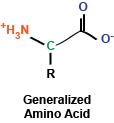
While I've written many posts describing signaling pathways and cellular phenomena of significant complexity, I'd like to use this post to take a step back and look at some fundamental building blocks, first turning to amino acids, the monomers that, when polymerized, make up polypeptides and proteins. At the most basic level, amino acids are really a rather simple chemical compound, consisting of a amino group, a carboxy group, a hydrogen, and a side chain, all sticking off a central carbon atom.  These amino acids are polymerized via their amino and carboxy chemical groups to create long, linear linkages.

Brief aside: In my chemistry class in undergrad, my TA helped us remember the order of the chemical bonds following polymerization by saying N-H, C-H, C-O, N-H, C-H, C-O, ...


You may remember briefly from any stint in chemistry class that a carbon atom that is covalently bound to four different chemical entities (in this case, a side chain, a hydrogen atom, a carboxyl group, and an amino group) can take two different conformations, depending on how these bonds are spatially oriented.  In the case of amino acids, the vast majority of amino acids found in our bodies and used to generate proteins are L stereoisomers.  This is a result of the amino acid synthesis machinery structure exclusively generating L amino acids.  There are exceptions, but we won't get into that.

As I mentioned, amino acids have a side chain: the part of the amino acid that endows it with its identity.  These side chains can be broken into a few groups that we will explore now:

The first set of side chains is the nonpolar, hydrophobic side chains.  The amino acids in this group include alanine, valine, leucine, isoleucine, glycine, methionine, and proline (structures shown to the right).  What you'll immediately notice is that these amino acid side chains are composed mostly of hydrogens and carbons.  Thus, these side chains do not contain polar covalent bonds and do not interact as readily with water (thus the term hydrophobic - they're "afraid" of water.  Some amino acids of note in this group are proline, which contains a ring structure that creates a "kink" in the amino acid, and methionine, which contains a sulfur atom.

The next set is composed of the aromatic side chains, which includes phenylalanine, tyrosine, and tryptophan.  These amino acids all contain an aromatic ring, which makes them relatively nonpolar; thus, they do not interact favorably with water.  These amino acids are involved in mediating protein protein interactions and are frequently found at the active sites of enzymes.

Next up: polar, uncharged side chains: asparagine, cysteine, glutamine, serine, and threonine.  These amino acids contain hydroxyl, sulfhydryl, or amide groups that mediate interactions with water, but they carry no net charge.  An amino acid of note in this family is cysteine, which can react with itself to form cystine, which is important in mediating the formation of disulfide bonds in protein structures.

We'll consider basic side chains next.  These amino acids consist of arginine, histidine, and lysine, which all carry a net positive charge in solution.  Of note, histidine is commonly found at the active site of enzymes to serve as a protein donor or acceptor.

Finally, we find acidic side chains: aspartate and glutamate.  In solution, these amino acids carry a negative charge and are considered acidic.

In the diagram at right, I've drawn up each of the amino acids along with their three-letter and one-letter codes.  These codes are frequently used to abbreviate long lists of amino acids.

Another brief aside:  Did you know that a single woman designated the amino acid abbreviations?  She chose letters that made sense for most amino acids (as you can see above).  For tryptophan, for example, she chose W because she envisioned saying tryptophan as twyptophan.  Kind of cool, huh?


As a summary, here are the amino acid abbreviations:

  • A, ala, alanine
  • C, cys, cysteine
  • D, asp, aspartate
  • E, glu, glutamate
  • F, phe, phenylalanine
  • G, gly, glycine
  • H, his, histidine
  • I, ile, isoleucine
  • K, lys, lysine
  • L, leu, leucine
  • M, met, methionine
  • N, asn, asparagine
  • P, pro, proline
  • Q, gln, glutamine
  • R, arg, arginine (think aRRRRginine)
  • S, ser, serine
  • T, thr, threonine
  • V, val, valine
  • W, trp, tryptophan (tWWWyptophan)
  • Y, tyr, tyrosine

So there you have it: 20 amino acids.  In addition to these amino acids, our bodies contain several more, including selenocysteine (identical to cysteine but containing selenium rather than sulfur) and ornithine (remember this from glycolysis?).  Amino acids can also undergo modifications: for instance, lysine residues can be acetylated.  More amino acids and their variants are always being discovered as well.

Now that we have the building blocks of proteins established, the next blog post will focus on how these amino acids can be combined (polymerized) into long structures that make up polypeptides and proteins.

LinkWithin

Related Posts with Thumbnails