Thursday, July 10, 2014

Optimizing a Western Blot

In the last post, we talked about the process of performing a Western blot, and a lot of the steps require optimization.  Exactly why is that?  How can you optimize these steps?

But why?!
Let's start from the beginning.  Why do we need to optimize Western blots at all?  Western blots rely on antibodies to detect proteins, and antibodies are created in animals.  This process involves purifying a protein (or a chemical), injecting the antigen (protein or chemical) into an animal, collecting that animal's serum, and purifying antibodies.  This process produces polyclonal antibodies, and the purification process can affect their specificity.  For many scientists, raising one's own antibodies is laborious and unnecessary, as many antibodies are commercially available.  If an antibody is not commercially available, one can purify a protein and send it to a company in order to produce an antibody.

The above process results in polyclonal antibodies.  Monoclonal antibodies can be derived by isolating B cells from stimulated animals, detecting which B cell population is producing the antibody against your antigen, and growing this B cell in culture.  Of course, more steps than this are involved, and the previous sentence is a very rough description.  Regardless, using this method, one can make virtually unlimited supplies of antibody.

Importantly, no two animals will react to an antigen in the same way - that's the beauty (and difficulty) of the immune system.  Thus, no two antibodies are really the same either - unless you have monoclonal antibodies.

To summarize:
  • Western blots rely on antibodies for detection
  • Creating antibodies is technically difficult and involves injecting an animal with an antigen
  • Polyclonal and monoclonal antibodies can be produced and they have different uses
  • Different animals produce different antibodies
  • Animals within the same species will make slightly different antibodies.
Using antibodies
Antibodies are used for many processes in biomedical science: Western blots, immunofluorescence, ELISA, immunoprecipitation...  If an antibody works (or doesn't work) for any of these processes, it may (or may not) work for another.  Prior to use, antibodies must be tested and optimized.

The steps that require optimization
Let's review the steps in performing a Western blot that require optimization
  • Blocking agent
  • Detergent concentration
  • Antibody concentration
  • Incubation times and temperatures
  • Secondary antibody concentrations, incubation times and temperatures
  • Exposure time

We'll go through these one at a time.

The blocking agent
In Western blotting, two blocking agents are typically used: non-fat milk or bovine serum albumin (BSA).  Typically, milk is used because it is cheaper than BSA.  Typical concentrations of milk range from 5-10% (weight by volunme).  However, for antibodies directed against phosphoproteins, BSA must be used because these antibodies will be neutralized by milk.

In addition to which blocking agent to use, one can also optimize the amount of time for blocking and the temperature.

In my personal experience, 5% milk in 2% TBST (Tris-buffered saline with Tween) for 1 hour at room temperature does the trick.  If you're in a rush, 30 minutes at 37 degrees can also work.

Detergent concentration
In Western blotting detergents are used to remove excess antibody and to prevent background noise.  Generally, a low concentration of detergent is sufficient to clear antibodies binding non-specifically to a membrane.  For me, 2% Tween works well, and it can be diluted in Tris- or phosphate-buffered saline.  For antibodies with greater background noise, the concentration of Tween can be increased, or the number / length of washes can be increased.

Antibody concentration
Probably the best place to start with optimization with with how much antibody you're using.  Naturally, you want to use less antibody (they're expensive, after all!), but it's good to try a range of concentrations.  For me, 1 uL of antibody in 1 mL (1:1000) is a good starting point.  Some antibodies work at 1:20,000; others work at 1:100.  It entirely depends on the antibody, and the manufacturer should provide guidelines for what concentrations to try.

Also, keep in mind that the concentration of your antibody depends on the application:  one concentration for Western blotting may not work for immunofluorescence.

Incubation times and temperatures
Yet another step that requires optimization is how long and how hot to incubate your membranes.  Sometimes, antibodies work quickly and you cannot incubate for long periods of time because this will result in higher background.  However, other antibodies will require longer incubations, even overnight, in order to see any bands.  Generally, longer incubations are done at 4 degrees, while shorter incubations (up to a few hours) can be done at room temperature.  You'll never know exactly what incubation time to use until you try.

Secondary antibody concentrations and incubations
As addressed above for primary antibodies, the same should be done for secondary antibodies - sounds like a lot of work, no?  The good news it that secondary antibodies are generally "well-behaved."  For instance, one particular type of secondary antibody uses a concentration of 1:20,000 in milk or BSA for an hour at room temperature.  No optimization required if you know what the conditions are!  However, if you're working with a new secondary, especially if it's a new species, optimization is your best bet.

Exposure time
One of the last steps to optimize is how long to expose a blot before developing it.  Modern technologies have attempted to supplant this step: new chemiluminescent detectors have reduced our need to measure how long to expose our blots.  However, for those of us that can't afford this fancy new equipment, we may rely on the good old film and developer.  Using this methodology, the amount of time film is exposed to your completed blot will affect how dark your bands are, as well as how much background you.  Of course, you want to optimize this step so that your image is clear and not misleading.  Overexposing a blot can lead to bands that all look the same; in reality, they might not be if you were to expose your blot less.  Additionally, taking several exposures - at both short and long lengths - will give you a range from which to choose.

Conclusions and ideas
The above gives a general guide for how to optimize a Western blot, but by no means is it exhaustive.  You can also optimize your developing reagent, for instance.  The best bet is to try something and tweak as needed.  Western blots are truly an art, and they can be really, really frustrating.  However, a little bit of effort in optimization will save you a lot of headache down the road...

Sunday, June 29, 2014

How a Western blot works

One of the most important techniques in molecular biology is the Western blot, which is used to detect proteins in a sample.  Running a Western blot can be an intimidating experience, as there are multiple steps, and if care isn't taken at each of the steps, the end product can be reduced in quality.

Several sources exist that explain in more technical detail how to run a Western blot, including this article from the National Institutes of Health.  Below is a summary of the theory and basics of how to run a Western blot.

The Sample:  To run a Western blot, we need some kind of sample, which in most cases is a cellular lysate, which we want to probe for the presence (and possibly abundance) of a protein.

Cell lysates from tissue culture can be collected by removing growth medium, washing in a neutral solution (such as PBS) and adding a lysis buffer to break open the cells.  Additionally, tissues from mouse organs or even human samples can be ground into lysis buffer, or bead-beaten into small pieces.  The lysed cells will need to be broken up in order to load them into our Western blot and in order to develop a nice final product.

After cell lysis, we've got two things to do.  First, we need to figure out how much protein we've got in the sample.  Determining protein concentration is usually done with a Bradford assay, which is a colorimetric assay.  A future post will deal with the details of this assay.

Next we also have to destroy DNA in the sample, while reducing disulfide bonds in our proteins.  Typically, this is done with a buffer containing beta-mercaptoethanol (the stinky stuff!) and boiling.  Protein samples are usually very viscous due to DNA, and if boiling doesn't get rid of all of the viscosity, a syringe and needle can be used to mechanically shear the DNA.

Running the Gel:  Many labs now use pre-made acrylamide gels, but many make their own gels as well.  A future post will have to delve into the details of what types and concentrations of gels to use.  For now, let's assume that we have the right kind of gel, the right percent acrylamide, and we've received the gel from a company.

In order to estimate the size of your protein product, a molecular weight marker should be run alongside the sample.  These ladders are commercially available and allow for approximation of molecular weights.  Being able to tell the size of a protein is important - sometimes you can be surprised by what size your protein looks compared to what you had expected!  If a protein is running at a "weird" size, don't shrug it off - that could mean something important.  In addition to running your samples and molecular weight marker in the gel, you want to fill every empty lane with buffer.  This will aid in running the gel and prevent proteins in your sample from shifting to the empty side of the gel.

Polyacrylamide gels are typically run at about 100-200V for anywhere from 30 minutes to three hours, depending on the size of the protein and the resolution you would like.  But what's really going on when the gel is running?  The buffer used to lyse cells contains sodium dodecyl sulfate (SDS), which is a negatively charged molecule (and it's an irritant, so always be careful when working with it), and molecules of SDS cover the proteins in the sample.  Since SDS is charged, applying a current to a gel loaded with protein covered in SDS causes this protein to migrate through the gel.  Bigger proteins (or bulkier proteins in general) move through the gel more slowly, so they don't migrate through the gel as quickly.

When a voltage is applied to the gel, the proteins migrate through two phases of the gel: the stacking layer and the resolving layer.  The stacking layer orders the proteins by length, based on their charge (from the SDS) and the resolving layer then expands the distance between these proteins, resulting in a fully resolved (and readable) gel.

After running the gel, it's time to transfer!  Transferring a Western blot involves moving your resolved proteins in the polyacrylamide gel into a membrane, usually PVDF or nitrocellulose.  This process can be done in the old school manner - by capillary action.  To use capillary action, the polyacrylamide gel is placed below the membrane, which is then stacked with paper towels.  The paper towels absorb moisture and draw proteins from the gel into the membrane, where they are "stuck."  This process is usually done overnight; thus, to reduce the amount of time this transfer takes, most researchers use either a wet or semi-dry transfer apparatus to use a current to draw proteins from the acrylamide into the membrane.

Following transfer, it's time to start to probe for our protein of interest.  Here is where Western blotting becomes an art:  everyone seems to do this step differently, and how this step is done depends on the antibody as well.  Regardless, there are three general steps.  First, we must block the membrane with nonspecific proteins, which is usually done with non-fat milk or with bovine serum albumin fraction IV (BSA).  The membrane is incubated in a solution for a specified amount of time at a specified temperature (such as 4 degrees, overnight or 37 degrees for an hour).  This process coats the membrane and prevents your antibody from binding non-specifically.

Next up:  primary antibody.  The primary antibody is the expensive reagent you can purchase from a number of vendors.  Antibodies can be easy to use, or they can be difficult.  They can be raised in mice, rats, chickens, goats, and more.  They can be monoclonal or polyclonal as well.  Thus, there is a lot of variability in antibodies, and your experiences with an antibody may be completely different from any other antibody.  This is part of the art of Western blotting: it is necessary to try things and to optimize your protocol.  Your membrane will be incubated with primary antibody, again for a specified amount of time, at a specified temperature and at a specified concentration.

Following incubation with the primary antibody, the membrane is washed in a solution containing a low amount of detergent (such as Tween), and then it's time for the secondary antibody.  The secondary antibody binds to the primary antibody and also contains some sort of means of detection - fluorescence or horse radish peroxidase (HRP) activity, for instance.  By using a secondary antibody, we greatly increase the specificity of the assay - in order to detect our protein of interest, that protein must be bound by the primary and secondary antibody.  The same conditions for the secondary apply as for the primary - one must figure out the best conditions for the antibody given the needs of the assay.

Immediately after incubation with the secondary antibody is detection - when things get interesting.  For the purposes of keeping this post short (kind of!), we'll describe the old-school method of exposure the blot to film.  After washing the excess secondary antibody off the membrane, one way to detect our protein is to use reagents that emit light via the HRP activity of the secondary antibody.  The detection reagent is added to the membrane for a short period of time and then removed.  Then, the membrane is moved to a dark room where the membrane is exposed to film for a specified amount of time.

After exposure, we need to develop the Western blot, by running the film through a developer.  Here's where we obtain our final product - a film with lines and smudges indicating (hopefully!) that our protein is where we hope it is.  At this point, we will know how much more optimization we need to do.

As you may have noticed, there is a lot of optimization of Western blots.  Below is a short list of steps that can be optimized:

  1. Running conditions of the polyacrylamide gel (percent polyacrylamide, voltage)
  2. Type of membrane - PVDF versus nitrocellulose
  3. Transfer conditions - wet, semi-dry, voltages, times
  4. Blocking conditions, times, and temperatures
  5. Membrane wash components - amount of detergent, number of washes
  6. Primary antibody incubation conditions  - times, concentrations, and temperatures
  7. Secondary antibody incubation conditions - times, concentrations, and temperatures
  8. Detection method
  9. Exposure time (if applicable)
Needless to say, Western blotting is an art.  The optimization steps must be done for every single antibody, which can be difficult, especially if the antibody is particularly difficult to work with.  Regardless, Western blotting is a very powerful and popular technique to detect proteins.

Saturday, June 21, 2014

Protein Structure II: Digging Deeper

In the previous post, we discussed the different levels of protein structure.  Here, we will consider more specific details of what makes up protein structure.

Previously, we talked about how secondary structure consists of alpha helices and beta sheets, but there are even more nuanced forms of secondary structure.  For example,  different types of alpha helices exist:  the 3.613 and 310 helices are different types that consist of 3.6 and 3 amino acids per turn of the helix, they also have different radii and lengths.

An interesting little tidbit about alpha helices is that there are certain amino acids that are more likely to be found in helices.  These include alanine, glutamine, leucine, and methionine.  In contrast, there are some amino acids that do not really fit into a helix: proline, glycine, tyrosine, and serine.  These amino acids have structural characteristics (such as proline's "kink") that tend to break helix structure.

Additionally, beta sheets have different properties depending on their orientations.  For example, with a parallel beta sheet, the chains of the protein are oriented in the same direction.  In contrast, anti-parallel beta sheets have chains that run in opposite directions.  See the illustration for a more visual explanation of this trait.

In addition to beta sheets and alpha helices, a few more "secondary structures" exist.  I put this in quotation marks because these types of structures are perhaps not classical structures - in that they're not alpha helices or beta sheets.  The first is the loop, which is a stretch of amino acids that makes a loop - rather self-explanatory!  These loops are important for protein structure because they allow for the creation of relatively compact proteins.  In particular, loops can be found in anti-parallel beta sheets, connecting the two beta strands to make the sheet.  Sometimes these structures are called hairpins because they are tight stretches of amino acids that hold a protein structure together.

The last secondary structural element we'll consider is the crossover loop, which is similar to the hairpin loop, but it connects portions of a protein at a longer distances.  These crossovers can be found especially in anti-parallel beta sheets, connecting the two strands such that they can be anti-parallel.

As mentioned, secondary structure consists of small tracts of protein structure, primarily formed of alpha helices and beta sheets.  These secondary structural elements can be organized into specific combinations called motifs.  Motifs are commonly-found structural organizations in proteins, such as zinc finger or coiled coil motifs.  In the case of a zinc finger motif, there are two beta strands and an alpha helix, making up a fold that looks something like a finger.  Within this motif is a zinc ion, hence its name.  Several proteins contain this motif, which is primarily involved in DNA- and RNA-binding.

Another level of structure is the domain, which is considered a module of a protein.  In general, domains have functions that can be separated from the protein as a whole.  Domains are typically large pieces of proteins (think of them as a swing set on a playground - the playground is for kids to play, and the swing set has its own specific function, to swing!).  An interesting aspect of domains is that they can be found in multiple proteins with similar functions.  For example, some proteins have kinase domains to help with phosphorylation; some have RNA-binding domains; and so on.  By detecting domains within a protein, we can infer its function, and this ability has been incredibly useful in predicting the function of new proteins.


Sunday, June 15, 2014

The Basics of Protein Structure Part 1: The Levels of Structure

In a previous post, we had explored the characteristics of the 20 amino acids that make up proteins.  These amino acids make up a diverse collection of molecules that can be strung together, making up proteins that have a multitude of functions within the cell.

Amino acids form a protein through the action of the ribosome, which we will discuss in a future post.  At this time, suffice it to say that the ribosome uses an elegant mechanism to read mRNA and translate it into the protein encoded by the mRNA by adding amino acids in a string.  As this string of amino acids is created, it begins to form a structure that will have functions within the cell.

Four levels of protein structure exist:
Primary (1o) structure:  The order of the amino acids is the primary structure.  Think of the primary structure as the alphabet of the amino acids: MGRYNVPL, for example.  The primary structure describes what order amino acids are in, and even though it might not seem like very much information, even the primary structure of a protein can provide a great deal of information in terms of its function and its potential 3-dimensional shape.

Secondary (2o) structure:  When amino acids are polymerized, they form local structures, which make up the secondary structure.  Think of secondary structure as the shape of a group of amino acids.  Two primary forms of secondary structure exist:  alpha helices and beta sheets.  Alpha helices result from the coiling of the amino acid string turning about itself.  In contrast, beta sheets are flatter and lack coiling.  There are several types of alpha helices and beta sheets, which we will save for a future post, and these different types of structures have important implications for overall shape of a protein.

Tertiary (3o) structure:  Protein structure gets exciting when you talk about tertiary structure, which can be described as the overall three-dimensional shape of a protein.  In general, the tertiary structure is the “final” form of a protein, although modifications on the protein, as well as interactions with other proteins can affect this structure.


Quaternary (4o) structure:  When proteins interact with each other, they can form complexes, which is the quaternary structure of these proteins.  You can think of this structure as the way proteins contact each other.  The bundling of proteins together can be between proteins of the same type (such as is the case with hemoglobin) or other types of proteins.

The drawing attempts to illustrate the concept of the different levels of protein structure.  Again, think of the primary structure as the order; the secondary structure as the local shape; the tertiary structure as the overall shape; and the quaternary structure as the way this tertiary structure of the protein interacts with other proteins.

How protein structure is established is a fascinating question and a field that is actively studied by prominent labs around the world.  Protein folding is the process of a chain of amino acids curling into its final shape, and how this process occurs is complex and not completely understood.  In general proteins fold depending on their environment (exposed to water or not, for example) and with the help of other proteins, called chaperones.  Protein chaperones help to establish a protein’s structure as well as maintain it during times of stress.  Further, modifications on proteins can change their structures, such as when p53, a protein that is involved in regulating many processes within the cell, is phosphorylated – its structure and, consequently, its function is altered slightly. 

The structure of proteins has fascinated scientists since we first learned about proteins.  Thousands of scientists around the world are still working to discover new structures and to learn how proteins fold.  The science behind protein folding has important implications in diseases such as Alzheimer’s, cancers, and infectious diseases.   

Sunday, June 8, 2014

The Basics: Prokaryotes versus Eukaryotes

Biology is an amazing complex topic, and this complexity is what makes it fun!

Before jumping into advanced topics, however, it is important to have a solid foundation of the basics, which we usually learn before algebra even.  As a quick review, let's talk about prokaryotes and eukaryotes, the two types of cells that should be familiar to most everyone.

The world of living cells is broken down into two major types of cells: prokaryotes and eukaryotes (also called procaryotes and eucaryotes, depending how you would like to spell).  Evolutionarily, we consider prokaryotes to be the ancestors of eukaryotes, which we will discuss further in the future.

Let's start with the prokaryotic cell and its defining characteristics:

  • Cell wall:  Prokaryotes have a cell wall, which are tough boundaries that enclose the cellular contents.  The cell wall is pretty tough, too - its composition gives it rigidity that keeps the prokaryotic cell's shape.  The cell wall is covered on the outside by the outer membrane and on the inside by the plasma membrane.  
  • Nucleoid:  DNA in prokaryotes isn't organized quite the same as in eukaryotes.  Rather than being organized in a distinct, membrane-bound portion of the cell, prokaryotes organize their DNA in what is considered the nucleoid.  You can think of the nucleoid as a membrane-less compartment where the organism's DNA is found.  Although in the diagram I have drawn a single linear DNA molecule, prokaryotes have a diversity of types of DNA, from single circular DNA to multiple linear and circular DNA molecules.
  • Cytoplasm:  The area within the cell wall and membranes is considered the cytoplasm - the fluid portion of the cell.  By no means is the cytoplasm empty space - it is filled with molecules and many events are taking place constantly within the cytoplasm.
Prokaryotes may seem "simple," but they certainly are not.  The above points are a simplification, but are the general characteristics of prokaryotes.

Eukaryotes can be considered more "complex," though certainly not better.  As with prokaryotes, the below characteristics are general and apply to most eukaryotic cells.
  • Nucleus:  Unlike prokaryotes, eukaryotes have a define space for their DNA - the nucleus.  The nucleus is surrounded by the nuclear membrane, which is surrounded by the endoplasmid reticulum (discussed next).  The nucleus is a hub of activity in the eukaryotic cell and by its enclosure in the nuclear membrane, distinct events can occur here and not in the cytoplasm.
  • Endoplasmic reticulum:  Surrounding the nucleus is the endoplasmic reticulum (ER), which is a compartment involved in moving molecules in and out of the nucleus, as well as regulating gene expression.  The ER can be broken down into the smooth and rough ER.  The rough ER is where proteins are made on the membrane.  In contrast, the smooth ER can be considered where pieces of the membrane are exchanged with the Golgi apparatus (next).
  • Golgi apparatus:  The Golig consists of a stack of membrane-bound vesicles, which are mainly involved in protein trafficking.  Here, a number of modifications can be made to proteins to target them to specific parts of the cell, such as the membrane or lysosomes.
  • Secretory vesicles:  Some molecules in the cell need to be sent outside the cell, and they can exit through secretory vesicles.  These vesicles often originate in the Golgi, after which they fuse with the plasma membrane and dump their contents in the extracellular space.
  • Lysosome:  The trash compactor of the cell, the lysosome is involved in recycling the contents of the cell.  Lysosomes have contents that break down molecules into their components so that the cell can reuse them.  Additionally, lysosomes can be used to destroy invading bacteria and to break down molecules that can be harmful to the cell.
  • Mitochondria:  "The powerhouse of the cell" - the mitochondria (singular mitochondrion) are involved in energy production, yes, but they also function is several other aspects, such as cell death.  Mitochondria themselves are like miniature cells within the eukaryotic cell, and they have their own DNA (mtDNA).  Mitochondria have many interesting aspects that will be discussed in a future post.
  • Plasma membrane:  Similar to prokaryotes, the plasma membrane in eukaryotes acts as a boundary, separating the cell from the outside.  Unlike prokaryotes, however, eukaryotes (generally) do not have a cell wall.  The plasma membrane is a very dynamic part of the cell - signaling, budding, engulfing.  
As mentioned, the above characteristics are an incredible simplification of prokaryotes and eukaryotes, but let this serve as a starting point into learning about each type of cell.  Both types have their own special attributes and many mysteries that remain to be solved.

Monday, November 14, 2011

Immunology II: Lymphoid Organs and Cells

As mentioned in the last post, several types of cells derive from lymphoid progenitor cells.  These cells are generated in the bone marrow in general, but only B cells mature there (hence the name B cells).  In contrast, T cells migrate to the thymus where they mature.  After full maturation of both B and T cells, they circulate in the blood system and then enter the peripheral lymphoid organs.  The central lymphoid organs are the bone marrow where the lymphocytes are generated, whereas the peripheral lymphoid organs are where T cells mature and where the adaptive immune responds to various stimuli.

The peripheral lymphoid organs
First, we will consider the components of the peripheral lymphoid system.  The lymph nodes are glands right near each armpit that is where fluid collects from the lymph system.  Lymph drains into the lymph nodes via lymphatic vessels and consists of the extracellular fluid filtered from the blood.  Thus, lymph is kind of a surveillance system for the body.  The afferent lymphatic vessels carry lymph and cells from infected tissues and drain into the lymph node.

The lymph node itself has a unique structure, illustrated to the right.  The follicles are where B lymphocytes set up shop, and T cells exist in paracortical areas (T-cell zones).  Germinal centers within the lymph node are where B cells proliferate after they have been stimulated by T cells.  Several additional tissues are organized similar to the lymph node drawn to the right, and this structure facilitates interaction between B and T cells.

The spleen is another peripheral lymphoid organ that mostly works to break down dead red blood cells.  This destruction occurs in the red pulp of the spleen, but the spleen also has white pulp where lymphocytes enter and exist within the spleen.  Within the white pulp is the periarteriolar lymphoid sheath (PALS) that contains T cells and a B-cell corona.

The digestive system is a major route for infection and has several gut-associated lymphoid tissues (GALT).  Some of these tissues include the tonsils, adenoids, and the appendix.  The intestine also has its own GALT, namely the Peyer's patches, which collect antigen directly from inside the intestine using multi-fenestrated (M) cells.

Similar to the digestive tract, the respiratory tract has its own lymphoid tissue, called the bronchial-associated lymphoid tissue (BALT).

Wednesday, November 9, 2011

The basics of immunology

Immunology scares me. I'm not ashamed to admit this fact. I find the topic intimidating and overwhelming, especially when I listen to talks given by prominent immunologists. The terminology is difficult, and the concepts seem very intertwined. I've always perceived that breaking into understanding immunology required a lot of work but that it would (and should) make sense... eventually.

 The next few blog posts are going to focus on immunology, not only because I need to learn this information, but also because it is fascinating and a challenging topic.

Components of the Immune System
All of the cells that comprise the immune system emerge from the bone marrow, where all of them originally come from and where some of them remain for maturation.  The cell type that gives rise to immune cells is the hematopoietic stem cell.  From this pluripotent state, the hematopoetic stem cell can then mature into a myeloid progenitor cell or a common lymphoid progenitor.  Myeloid progenitor cells can differentiate into several more cell types, including granulocyte and macrophage progenitors and megakaryocyte and erythrocyte progenitors.  The granulocyte and macrophage progenitors can then develop into neutrophils, eosinophils, basophils, mast cells, and macrophages.  Megakaryocyte and erythrocyte progenitors generate platelets upon maturation.

Hematopoietic stem cells can also develop into a common lymphoid progenitor, which consists of B cells, T cells, and NK cells.  These types of cells leave the bone marrow and migrate through the lymph nodes.  Dendritic cells also develop from lymphoid progenitor cells but mature in the bone marrow before entering the lymph node.

Basic functions of immune cells
  • Macrophages are a common cell type that mature from monocytes (from the myeloid progenitor cells originally).  Monocytes circulate in the blood and continuously differentiate into macrophages when they enter the body's tissues.  Once in the tissues, macrophages can be considered the garge trucks of the body:  they engulf the environment as well as other cells in the process of phagocytosis.  Thus, macrophages can function to neutralize harmful elements within the body.
  • Dendritic cells also mature from myeloid progenitor cells, and their main function is to process and display antigen that will then be readable by T lymphocytes.  This antigen display requires the presentation of co-stimulatory molecules, and when dendritic cells encounter a pathogen (or other foreign antigen), they mature and begin expressing these co-stimulatory molecules.
  • Mast cells differentiate in body tissues and are involved in mediating mucosal immunity.  They are most well-known for their role in allergic reactions.
  • Neutrophils are a type of granulocyte (so called because they have densely-staining and strange-shaped nuclei) that are involved in phagocytosis and increase in numbers upon an immune response.
  • Eosinophils respond to parasites.
  • Basophils may function similarly to mast cells.
  • B cells differentiate into plasma cells and function to secrete antibodies.
  • T cells destroy virus-infected cells and also function to activate other immune cells, such as B cells and macrophages.
  • NK cells are involved in innate immunity and destroy "weird-looking" cells, such as tumor cells or cells infected with viruses.
References for the interested:
Immunobiology. Janeway, Travers, Walport, Shlomchik.
Basic Concepts of Immunology and Neuroimmunology: Basic Immunology

Wednesday, November 2, 2011

Cancer and Oncogenes

Cancer is a diverse group of diseases with one common characteristic: unchecked cellular replication.  Via several potential mechanisms, cancer cells are able to avoid all of the checkpoints involved in cell growth and division, thus enabling them to divide more frequently or indefinitely.  Many events can lead to the development of a cancer cell, including inheritance of mutated DNA or the activity of a carcinogen, or a chemical agent that leads to the development of cancer.

Gene expression is often deregulated in cancer cells such that some genes are overexpressed, while others are underexpressed.  Genes that can be mutated to lead to an upregulation of activity and lead to the development of a cancer cell are termed proto-oncogenes.  When these proto-oncogenes are actually mutated, they are considered oncogenes.


An oncogene is often a gene involved in regulating cell division and drive the cell cycle.  When they are overexpressed, such as during cancer, they can push the cell to divide more frequently and, with further mutations, transform the cell such that it divides without restriction.

Oncogenes were first discovered in viruses, specifically in Rous Sarcoma Virus (RSV), a retrovirus that encodes a homologue to cellular src kinase (the viral form called v-src).  Tumors in birds caused by RSV are the result of v-src causing unregulated cellular proliferation.  Large amounts of research into this area has identified cellular src kinase as a proto-oncogene that, when mutated to become constitutively active, becomes an oncogene and can drive cancer development.  Interestingly, viruses have highlighted a number of cellular oncogenes and pathways that are improperly regulated in cancer.  Over 20 viral oncogenes have been identified to date.

Cellular proto-oncogenes (the genes before they become oncogenes) can promote cellular proliferation and the development of cancer in several ways.  One of these ways is to be overexpressed and function when the gene product really shouldn't function.  This is the case with proteins such as myc and growth factor receptors.  With overexpression of these proteins, there is the potential for amplified signaling through these pathways that can push the cell to divide more than it normally does, leading to the development of cancer.  An additional mechanism whereby a proto-oncogene can become an oncogene is via mutation that leads to improper regulation, such as constitutive activity.  A classical example of this type of phenomenon is via Ras, which when mutated is constitutively active and cannot hydrolyze an attached GTP to inactivate.  Thus, Ras remains active and cannot be "turned off."  This constant activity of Ras results in  signal transduction to the nucleus of the cell and pushes the cell to divide through transcription of several genes involved in cell division.


Monday, October 17, 2011

The Trp Operon

The last post about an operon (the lac operon) is the most viewed post on this blog, so I thought that it might be helpful to follow this up with another operon, this time concentrating on the trp (tryptophan) operon.  This operon is another really elegant example of transcriptional regulation in E.coli and the mechanism is pretty cool.

Amino acids are essential for life (see the last post on their composition!) and cells synthesize amino acids using a variety of enzymes.  When nutrients are plentiful, such as E.coli would encounter in nutrient broth in the laboratory setting, cells no longer need to waste energy producing biosynthetic enzymes when they can utilize nutrients already in excess.  The trp operon contains several enzymes that are coordinately regulated and involved in the production of tryptophan.  When tryptophan is present in the cell's environment, it doesn't need to make any of these enzymes, but if the cell needs tryptophan, these enzymes are transcribed and shortly thereafter translated.  Control of this operon, thus, controls how much energy the cell is going to put into making tryptophan.

Similar to the lac operon, the trp operon contains an operator (O) sequence, within the promoter sequence, where an operator binds and prevents transcription.  In the presence of tryptophan, the operator binds the promoter and prevents RNA polymerase from transcribing genes.  In the absence of tryptophan, however, transcription occurs at a basal rate.  Sounds simple enough, right?  Let's take it a step further and consider...

Attenuation
An important concept in gene regulation is that of attenuation, which is fine-tuning of gene expression.  You might think that attenuation is mediated by protein factors that bind the DNA and affect gene expression; however, attenuation of the trp operon is a little different and, instead, depends on mRNA structure to modulate gene expression.

Before moving forward, let's look at the trp operon (diagrammed to the right).  Briefly,t here are four regions, and these four regions have differing levels of complementarity to each other.  Thus, when the DNA is transcribed into mRNA, the mRNA folds into all kinds of shapes and the regions of the trp operon fold on each other.

After transcription of the entire trp operon (we're dealing with mRNA from this point forward), the next event is translation of this mRNA into protein.  In bacteria, it's important to note that transcription and translation occur simultaneously, so as soon as we have a transcript in a bacterial cell, it's being translated.  The trp transcript contains two critical tryptophan codons immediately before region 1, so in order to synthesize the enzymatic machinery to make tryptophan, the cell must use a few residues to translate the protein.

In the presence of high amounts of tryptophan within the cell, the ribosome plows through these two tryptophan codons, adding in the appropriate amino acids, and continuing through region 1 of the mRNA.  This results in region 1 and 2 mRNA sequences binding together, and then regions 3 and 4 bind together as well.  This interaction between regions 3 and 4 results in the creation of a transcription-termination hairpin, basically a structure in the mRNA that kicks out RNA polymerase and prevents further transcription of the mRNA.  Thus, transcription (and then translation) are stopped because

In the absence of tryptophan, however, the ribosome cannot quickly add tryptophan during the translation process and it stalls before region 1.  This results in the folding of the mRNA such that regions 2 and 3 bind to each other.  When this structure forms, no transcriptional termination hairpin is formed, and mRNA synthesis continues.  Thus, the entire mRNA sequence for the trp operon is made and can be translated into enzymes that will synthesize tryptophan.

In summary:
Lots of tryptophan: Ribosome zooms through the mRNA, regions 1 & 2 and 3 & 4 bind (in pairs) and create a termination hairpin
End result: Transcription terminates and tryptophan synthetic enzymes not created (cell saves energy!)

Lack of tryptophan: Ribosome stalls immediately before region 1, regions 2 and 3 bind each other, no termination hairpin is formed
End result: Transcription continues and biosynthetic enzymes are eventually synthesized

This scheme is similar for other operons encoding amino acid biosynthetic enzymes (in bacteria, that is).  The trp operon is an elegant scheme to finely-tune transcription via mRNA structure to prevent the cell from wasting energy.

Thursday, October 13, 2011

Amino Acids: The Building Blocks of Proteins

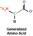
While I've written many posts describing signaling pathways and cellular phenomena of significant complexity, I'd like to use this post to take a step back and look at some fundamental building blocks, first turning to amino acids, the monomers that, when polymerized, make up polypeptides and proteins. At the most basic level, amino acids are really a rather simple chemical compound, consisting of a amino group, a carboxy group, a hydrogen, and a side chain, all sticking off a central carbon atom.  These amino acids are polymerized via their amino and carboxy chemical groups to create long, linear linkages.

Brief aside: In my chemistry class in undergrad, my TA helped us remember the order of the chemical bonds following polymerization by saying N-H, C-H, C-O, N-H, C-H, C-O, ...


You may remember briefly from any stint in chemistry class that a carbon atom that is covalently bound to four different chemical entities (in this case, a side chain, a hydrogen atom, a carboxyl group, and an amino group) can take two different conformations, depending on how these bonds are spatially oriented.  In the case of amino acids, the vast majority of amino acids found in our bodies and used to generate proteins are L stereoisomers.  This is a result of the amino acid synthesis machinery structure exclusively generating L amino acids.  There are exceptions, but we won't get into that.

As I mentioned, amino acids have a side chain: the part of the amino acid that endows it with its identity.  These side chains can be broken into a few groups that we will explore now:

The first set of side chains is the nonpolar, hydrophobic side chains.  The amino acids in this group include alanine, valine, leucine, isoleucine, glycine, methionine, and proline (structures shown to the right).  What you'll immediately notice is that these amino acid side chains are composed mostly of hydrogens and carbons.  Thus, these side chains do not contain polar covalent bonds and do not interact as readily with water (thus the term hydrophobic - they're "afraid" of water.  Some amino acids of note in this group are proline, which contains a ring structure that creates a "kink" in the amino acid, and methionine, which contains a sulfur atom.

The next set is composed of the aromatic side chains, which includes phenylalanine, tyrosine, and tryptophan.  These amino acids all contain an aromatic ring, which makes them relatively nonpolar; thus, they do not interact favorably with water.  These amino acids are involved in mediating protein protein interactions and are frequently found at the active sites of enzymes.

Next up: polar, uncharged side chains: asparagine, cysteine, glutamine, serine, and threonine.  These amino acids contain hydroxyl, sulfhydryl, or amide groups that mediate interactions with water, but they carry no net charge.  An amino acid of note in this family is cysteine, which can react with itself to form cystine, which is important in mediating the formation of disulfide bonds in protein structures.

We'll consider basic side chains next.  These amino acids consist of arginine, histidine, and lysine, which all carry a net positive charge in solution.  Of note, histidine is commonly found at the active site of enzymes to serve as a protein donor or acceptor.

Finally, we find acidic side chains: aspartate and glutamate.  In solution, these amino acids carry a negative charge and are considered acidic.

In the diagram at right, I've drawn up each of the amino acids along with their three-letter and one-letter codes.  These codes are frequently used to abbreviate long lists of amino acids.

Another brief aside:  Did you know that a single woman designated the amino acid abbreviations?  She chose letters that made sense for most amino acids (as you can see above).  For tryptophan, for example, she chose W because she envisioned saying tryptophan as twyptophan.  Kind of cool, huh?


As a summary, here are the amino acid abbreviations:

  • A, ala, alanine
  • C, cys, cysteine
  • D, asp, aspartate
  • E, glu, glutamate
  • F, phe, phenylalanine
  • G, gly, glycine
  • H, his, histidine
  • I, ile, isoleucine
  • K, lys, lysine
  • L, leu, leucine
  • M, met, methionine
  • N, asn, asparagine
  • P, pro, proline
  • Q, gln, glutamine
  • R, arg, arginine (think aRRRRginine)
  • S, ser, serine
  • T, thr, threonine
  • V, val, valine
  • W, trp, tryptophan (tWWWyptophan)
  • Y, tyr, tyrosine

So there you have it: 20 amino acids.  In addition to these amino acids, our bodies contain several more, including selenocysteine (identical to cysteine but containing selenium rather than sulfur) and ornithine (remember this from glycolysis?).  Amino acids can also undergo modifications: for instance, lysine residues can be acetylated.  More amino acids and their variants are always being discovered as well.

Now that we have the building blocks of proteins established, the next blog post will focus on how these amino acids can be combined (polymerized) into long structures that make up polypeptides and proteins.

Friday, September 30, 2011

Android Applications for Scientists (and other people too!)

If you're like me, you like to use your phone for its capabilities, including for work, and because it's shiny and you paid a lot of money for it.

I use an Android phone (no thank you, Apple) and am always interested in an application that could help me with my research or studying.  Unfortunately, the Android marketplace is cluttered with irrelevant applications, making finding useful applications difficult.

The following applications are presented as a summary of what I use.  Certainly there are more out there (please tell me!) and more are created every day, and I look forward to using these applications in the future:

Astrid Tasks:  Every morning as I'm walking into the building, I fill out my task list of things to do for the day.  I don't need anything fancy - I just need an application that is quick, easy, and simple.  Astrid is fantastic for putting together this list and for prioritizing my experiments and work.  Plus, it's got a handy widget (if you're into widgets, that is).  I would highly recommend this application for those who like to keep lists.

GoogleDocs: If you use Google for composing, sharing, or viewing documents, this application is fantastic because it will sync your computer and your phone to view the same documents.  Additionally, if you've got a big enough screen, it's not too horrible to actually compose in these documents.  I don't use this too often, but it's convenient  when I've got a document to get on my phone and my Dropbox happens to be full (see below).

Doodle:  Have you ever tried to schedule a meeting with faculty?  How about with multiple faculty?  Needless to say, it is a nightmare: herding cats as many would say.  Doodle attempts to make this a little bit easier by creating a spreadsheet which participants can then check off for their ability.  That one magical time spot that everyone checks is then the meeting time.  Doodle comes with several options for creating an event and then adding participants.  In my department, these things are really the best way to make sure that a meeting is really going to happen.

Dropbox:  If you're not already on Dropbox, seriously, sign up for it.  This program is the most useful thing I have every used.  Once installed on a few computers and on your phone, a folder is created - your Dropbox.  It acts just like a normal system folder and can be manipulated just like one too.  The great thing is that anything that is in your Dropbox can be accessed on any computer that has Dropbox installed and you can also access your files online.  This is so much better than carrying around a flash drive, and the syncing is instantaneous.  I could not recommend this program more.

handyCalc:  Usually, I'll use my crappy old calculator from high school to do the simple arithmetic needed for my experiments and notebook.  When I'm at my computer and can't find my calculator, I use handyCalc, mainly because I find it much easier to use than the standard calculator that came with my phone.  There are multiple iterations of calculators out there - from simple to mind-bogglingly complex.  This program will solve equations, create graphs, and perform simple addition and subtraction.  It works and I like it.

LinkedIn:  At a recent conference, I learned the importance of LinkedIn.  The seminar speaker asked everyone in the room to raise their hand if they were a member of LinkedIn.  I was the only person not to raise my hand.  LinkedIn is a Facebook for professionals - it can be helpful if you're looking for a job or want to make some contacts to look for a job in the future.  Being able to use it on my phone is convenient, too.

Pulse:  I like to read the news on my phone when I can't access a computer.  The best app I've found for this is the Pulse reader app.  Using this program, you can view tiles containing the top headlines from various websites, including numerous science-slanting websites.  The handy swipe-to-change-story feature is nice, and the entire interface is easy-to-use.  I also like using this while I'm walking because it's easy to pull up a short story that I can finish quickly and make myself feel like I accomplished something.

QuickOffice:  I'm not a big fan of paid applications - I am a poor graduate student after all. QuickOffice came preinstalled with my phone, and I must say that I am quite impressed with it.  It has the same features as the Microsoft Office suite (I can even view my PowerPoint slides on my phone!) and is really easy to use.  If you're not up for shelling out $15 (!), you can always opt for the Google Docs app, which I think is almost just as good, and you can't beat the price...

Google Reader: I use Google Reader religiously for keeping up on journal articles.  This handy little app presents my RSS feed conveniently and in my pocket so that I can keep up on my papers that I need to read.  Since I can't access the actual papers on my phone, I star the articles of interest and download on my computer when I'm connected to my university's network.

The Weather Channel:  I work in a lab with no windows.  Most of the time, I can't tell is there's a tornado outside or a beautiful sunny day.  Sometimes, that's for the best because then I'm not tempted to leave my benchtop and wander outside.  When I do have to go outside, however, it's nice to know the weather, and the Weather Channel app is convenient for quickly checking the weather.  Tons of functionality are included, such as animated weather maps.  There are a lot of weather applications out there, but I find this to be the most useful.

WTFSIMFD:  Hands down, my favorite food app.  WTFSIMFD provides you with a recipes that you should eat.  The app has a potty mouth, but it's amusing and endearing at the same time.  It also gives great recipes, you know, for when you're not in the lab or studying...

Just a roundup of what I find useful as a student and a scientist.  Maybe a followup post will be necessary when I find even more useful apps...

Saturday, September 24, 2011

Mendelian Genetics Part I

Gregor Mendel was an accomplished scientist, though he never would know this during his time. His studies of pea plants (as well as other organisms) laid the groundwork for the genetic breakthroughs that would come after his death. Mendel was a Augustinian monk that had ample time to experiment with his pea plants. The principles he was able to extract from his studies are the basics taught in high schools around the world.

 Mendel studied peas, but not just any peas. His peas were true-breeding, meaning that they self-fertilized and produced essentially clones of themselves.  Over the years of study, he bred plants with specific characteristics and then crossed different varieties of pea plants to test what their offspring would look like.

Importantly, Mendel considered discontinuous, contrasting traits: he only considered the traits that he could observe (such as color), came in a limited number of forms (green or yellow) and were easily distinguishable.  For these monohybrid crosses, Mendel considered seven different traits: seed shape (round versus wrinkled), seed color (yellow versus green), pod shape (full versus constricted), pod color (yellow versus green), flower color (violet versus white), flower position (axial versus terminal), and stem height (tall versus short).  Also important to Mendel's work was his use of mathematics and statistics to estimate the probabilities of a certain type of plant emerging from a certain type of pea plant cross.

The monohybrid cross is the consideration of one particular trait at a time.  For example, if looking at pea pod color, one might cross a yellow and a green pea plant and then determine how many of the offspring had yellow or green pea pods.  This type of cross is illustrated to the right in what's called a Punnett Square.  A Punnett square is a way of organizing the different traits expressed by the individuals being crossed.  The capitalized letters are the dominant traits, while the lower-cased letters are recessive.  Vertical columns traditionally represent females; horizontal, males.  Using the Punnett square, we can look at all the possible offspring that can emerge from a cross, which can then be used to determine probabilities associated with the offspring's traits.

From these monohybrid crosses, Mendel was able to make a few conclusions.  When he crossed two pea plants of distinct traits (the P1 generation, true-breeding variety) to produce progeny (the F1 generation) and then used the progeny to generate more progeny (the F2 generation), he found that the two parental traits were still present and unchanged.  This led to the hypothesis that each parent contributed equally to the inheritance of the "genetic determinants," which would have more technical and molecular definitions in the distant future.  These determinants were separated and segregated randomly to make gametes and produce the next generation of pea plants.

Mendel's principles from the monohybrid crosses can be summarized as follows:

  1. Hereditary determinants (unit factors) control traits that are in pairs in an individual.
  2. When two dissimilar unit factors for a trait are combined in one organism, one factor is dominant to the other recessive factor.
  3. When gametes are formed, the pair of unit factors separate and are equally likely to be separated into a gamete: the principle of segregation
In the next post, we'll examine dihybrid crosses and some more of the interesting findings from, of all things, pea plants.


Thursday, September 22, 2011

MAP Kinase Signaling

Cells exist in a very dynamic environment, not only on the inside of the cell membrane, but also on the outside.  Thus, cells must be able to interact with their outside environment and respond to stimuli appropriately.  One of the several signaling pathways involved in communication from the outside of the cell inward is the (very general) MAP kinase (MAPK) signaling pathway.  The cell uses the MAPK signaling pathway for several reasons, primarily to amplify signals.  Additionally, aberrant MAPK signaling is implicated in several types of cancers.

First, MAPK signifies mitogen activated protein kinase - a fancy word for a protein that responds to mitogens, or a molecule that stimulates mitosis.  Proteins of the MAPK family were discovered in 1989 in yeast, with ERK1 being the first mammalian MAPK signaling protein, involved in insulin signaling pathways (at the time at least...).

The MAPK signaling pathway is often called a cascade because the components in the pathway amplify a signal within the cell.  The three major components are:
  • MAPKKK / MEKK / MAP3K: The MAP kinase kinase kinase.  This protein phosphorylates MAP kinase kinase.  Several forms are found within the cell and their abundance is lower than that of MAPKK or MAPK; thus, MAPKKK isn't as involved in the amplification of signals.
  • MAPKK / MEK / MAP2K: The MAP kinase kinase.  MAPKK phosphorylates MAPK and is highly abundant within the cell, but its only substrate is MAPK.  Just a few phosphorylation events on MAPKK result in significant activation of MAPK.
  • MAPK / ERK: The MAP kinase.  MAPK is also abundant in the cell and has diverse substrates, including itself.
There are several pathways that involve MAPK signaling.  Two of the important ones are:
  1. The ERK pathway: growth factor stimulation of cell surface receptors (via receptor tyrosine kinases [RTKs]) causes activation of Ras, which activates Raf (MAPKKK) to activate MEK (MAPKK) and then ERK (MAPK).  ERK's several targets include Elk and Ets, which control cellular proliferation.
  2. The JNK pathway: Stress on the cell results in activation of several proteins, including Rho, which goes on to activate MEKK, then MEK, and JNK.  JNK's major targets include c-Jun, ATF2, and Elk1, which control proliferation and apoptosis.
One important consideration is the specificity of MAPK signaling.  Specificity can be achieved in several manners: protein components can only "fit" other specific protein components.  Additionally, the spacial organization of a cell can affect specificity, and this type of specificity is often controlled by scaffold proteins.  

MAPK signaling is essential for many cellular processes, including proliferation, apoptosis, embryonic development, and cancer progression.  Many intricacies of the pathways are still being worked out and will provide significant insight in the future.

Thursday, September 1, 2011

The Basics of Protein Translation

Proteins, a major constituent of the cell, have many diverse functions and are classically considered to be the "work horses" of the cell.  Additional molecules, such as RNAs and lipids, have shown importance in signaling and catalyzing chemical reactions; however, proteins remain an important part in the life of a cell.

Before proteins can perform their evolved functions, they must be synthesized within the cell.  The process of synthesizing a protein is critically important to the cell and, thus, is an energy-intensive process.  

The basic building blocks of a protein are amino acids, which come in twenty (and more) flavors.  These amino acids have different properties that afford proteins different structures and functions when the amino acids are polymerized together in distinct orders.  These amino acids can have distinct signaling roles when they exist as monomers as well (see this paper by Nobukuni et al for an example).  

Monomeric amino acids in the cellular environment do not randomly polymerize to form proteins.  In the first of several steps, tRNAs are charged: they are covalently linked to amino acids via aminoacyl tRNA synthetases.  These synthetases hold the very important role of attaching the appropriate amino acid to the appropriate tRNA.  Because inappropriate charging of tRNAs would lead to misincorporation of amino acids into a protein chain (wasting energy or leading to even bigger problems for the cell), sythetases are very specific.  In fact, some synthetases have an editing site, where they will catalyze the removal of incorrectly placed amino acids.

After tRNAs are charged with their appropriate amino acids, they are ready for interaction with the ribosome.  Ribosomes are large, complex molecules that merit their own post.  Briefly, ribosomes are composed of RNA and protein and are made of two distinct complexes: the large and small subunits (depending on the origin of the ribosome, the subunits have different sedimentation coefficients, so you might see 30S and 50S for bacteria or 60S and 40S for eukaryotes, for example).  Ribosomes catalyze the polymerization of amino acids into proteins.

In the first step of ribosome-mediated protein production, the small subunit of the ribosome combined with a tRNA for methionine (the amino acid that begins the protein chain) scans along the transcribed mRNA until it encounters a start site (ATG codon).  Here, the complex stops, and the charged tRNA with its amino acid comes into contact with the peptidyl transferase site (P) on the ribosome.  eIF2 (eukaryotic initiation factor 2), which was along for the ride, hydrolyzes GTP to GDP at this point such that the ribosome stops at the appropriate codon.

Next, the large subunit of the ribosome binds the small subunit, making a full ribosome that is ready for catalysis.

The tRNA that lines up with the mRNA's next codon then binds in the acceptor site (A), along with the help of eEF-1 (eukaryotic elongation factor-1), which hydrolyzes GTP to GDP.  At this point, the ribosome goes into action: using the peptidyl transferase center (PTC), it catalyzes the covalent linkage of the first and second amino acids.

The entire ribosome now moves along the mRNA in a process called translocation, which requires the help of EF-2 (and GTP hydrolysis).  The first tRNA is moved into the exit site (E), and the second tRNA moves into the P site, while the A site is open for another aminoacyl-tRNA.

The process repeats until the ribosome encounters a stop codon.  At this point, termination factors (TFs), which have structures similar to tRNAs and bind mRNAs but do not have amino acids, enter the acceptor site of the ribosome.  The ribosome then catalyzes the addition of water to the end of the amino acid chain, releasing it from the peptidyl transferase center and allowing it to leave the ribosome and begin folding into its native conformation.

While there are several details that I may have seemingly glazed over, this post should give a broad, simplified overview of translation.  Future posts will address the many details involved.

Saturday, August 27, 2011

The Many Functions of Ubiquitin

Ubiquitin (abbreviated as Ub) is described as a small, frequently-encountered molecule involved in the proteosomal degradation of proteins.  In reality, the addition of ubiquitin moieties to proteins can serve several functions and is now recognized as a common post-translational modification (PTM).

The classical ubiquitin pathway that first comes to mind is that of proteosomal degradation.  In this pathway, illustrated in the accompanying figure, ubiquitin is first "activated" by binding covalently to a cysteine residue on E1, the ubiquitin-activating enzyme, which requires the energy of ATP.  Then ubiquitin is transfered to a cysteine residue on E2, the ubiquitin-conjugating enzyme (no ATP is required here).  Finally, E2 binds to a scaffold called E3, the ubiquitin ligase, which also contains the target protein - illustrated in pink.  This target protein is then covalently linked to ubiquitin via a lysine residue.  Several rounds of this pathway then result in linear polyubiquitination of proteins, which is then recognized by other components in the cell that result in its recruitment to the proteasome and subsequent degradation (a topic of future posts).  The end result is that the target protein is broken down into small seven-to-eight amino acid pieces.

As mentioned above, there are three proteins involved in the attachment of ubiquitin to target proteins, E1, E2, and E3.  In the cell, E1s are the least plentiful (only one has been discovered thus far), while the human genome codes for tens of E2.  Several more E3 ligases have been discovered (and more every day).

Importantly, ubiquitination does not exclusively function in proteosome-mediated degradation of proteins.  Monoubiquitination, or the attachment of a single ubiquitin moiety to a protein, has been shown to have diverse effects on target proteins (see listbelow). Additionally, multiple monoubiquitination events can occur on a single protein, as can branched polyubiquitination.

Ubiquitin is not the sole small protein modifier.  Several molecules similar to ubiquitin have been described and exhibit diverse functions in cellular signaling (topics of future posts).  These proteins include SUMO (small ubiquitin-like modifier), NEDD, FATIO, and FUBI.  Needless to say, all of these proteins have some of the most adorable names in science.

The following list is a short compilation of ubiquitin functions, and, as we all know very well, no hard-and-fast rules exist for ubiquitin modifications (or science in general).
  • p53 ubiquitination by MDM2: cell cycle regulation, apoptosis
  • HIF1 ubiquitination by VHL: hypoxic response
  • Caspase 3 and 7 ubiquitination by XIAP: apoptosis
  • PCNA: DNA repair
  • RNA polymerase II: transcriptional regulation
  • H2AX and H2A: transcriptional regulation, protein recruitment
Of course, there are more proteins that are modified by ubiquitin, but I hope that the above table illustrates the diversity of the modification.

Interestingly, ubiquitin modifications are not forever: de-ubiquitinating enzymes (DUBs) remove ubiquitin moieties from tagged proteins.  Thus, the reversible ubiquination represents a dynamic cellular process.

Some cool (and important) papers and links:
Principles of ubiquitin and SUMO modifications in DNA repair
Non-traditional Functions of Ubiquitin and Ubiquitin-binding Proteins
The Ubiquitin System
Nonproteolytic Functions of Ubiquitin in Cell Signaling
Ubiquitin Function and Variety

Tuesday, June 22, 2010

Initiation of Eukaryotic Transcription IV

Changing Up the Chromatin
All of the factors that affect transcription that have been described are involved in changing the ability of RNA polymerase and transcription factors to bind to and initiate DNA transcription.   An additional method of modulating transcription is via changes to the chromatin (see writings on that here).  Histones and other proteins are able to change the packaging of DNA and, thereby, affect transcription.  The compaction of the DNA directly affects the accessibility of it to RNA polymerase and transcription factors.  Euchromatin is the active form of chromatin that is not fully compacted; in contrast heterochromatin is generally not active or accessible.  The presence of histone H1 (the linker histone, and, let’s be honest, everyone’s favorite histone) also affects the compaction of the chromatin.

How exactly do histones affect transcription?  Put simply, they prevent other proteins from binding the DNA, especially the DNA that is facing the histone core.  Further, DNA is wrapped around histones, which changes the structure of the DNA.  Such distortions can affect binding sites and preclude transcription factor binding.  If the transcription factors and RNA polymerase are to bind to the DNA, the chromatin must be unraveled and the DNA must become accessible.  One way to do this is to move the histones out of the way, opening up binding sites.  Histones are incredibly dynamic and move around on the DNA frequently, wrapping and unwrapping different sequences.  With the fluctuations of the chromatin, transcription factors can bind while the histones “breathe.”

An additional mechanism to allow access to the DNA is via histone modifications on the N-terminal domains.  The N-termini of the histones contain lysine groups that affect DNA-histone interactions.  Therefore, affecting the charged residues via acetylation or methylation, for example, will change the interactions between the histones and DNA.  Acetylation of a histone tail effectively neutralizes its charge such that its structure is altered and no longer binds DNA as tightly.  Modifications on histones can also form binding sites for transcription sites.  Chromodomains bind to methylated lysine residues, while bromodomains bind acetylated lysines.  There are a number of different modifications that affect transcription, and new effects are still being elucidated.

-          Acetylation of lysine / arginine: transcription induction
-          Phosphorylation of serine, threonine, or tyrosine: transcription induction; chromatin compaction
-          Methylation of arginine: transcription induction
-          Methylation of lysine: gene silencing
-          Ubiquitination of H2A and H2B: degradation; transcription; growth regulation
-          ADP-ribosylation: histone repelled from DNA at sites of repair

LinkWithin

Related Posts with Thumbnails